


Breasts are the most prominent feature of a Woman’s body (Women Chest Muscles Anatomy), consisting of adipose tissue, mammary glands, milk ducts, and retaining ligaments. Having well-shaped breasts strengthens women’s sense of self-confidence and at the same time is considered one of the criteria for beauty for women. Aside from the strong chest muscles increasing the overall strength and efficiency of your body, the exercise of these muscles plays an undeniable role in improving the overall appearance of the upper body. In this article, we are going to examine in detail the Anatomy of women’s chest Muscles.
Women’s Chest Muscles
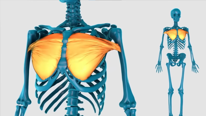
One of the superficial muscles in the thoracic area is the pectoral muscle, which consists of two pectoral parts, the major and minor. The major pectoral muscle itself consists of two parts the upper cords of the clavicular head and the lower cords or the winged head. The muscle attaches to the sternum and scapula on one side and the anterior part of the arm bone on the other. It also consists of two parts, the sternum and the clavicle. It is activated in the movements of the shoulder joint of the large pectoral muscles and in the movements of the shoulder of the small pectoral muscles. Knowing the anatomy of women’s breast muscles has a huge impact on breast care.
The large pectoral muscle is connected to the arm bone, but the small pectoral muscle is connected to the shoulder bones. Although this part of the body contains only two main muscles, it is one of the largest muscles in the body and requires a continuous and appropriate training program to strengthen and grow fully. In addition, the position of the anterior deltoid muscles, especially the junction of the pectoral and shoulder muscles in the upper and outer part of the chest and the front tooth muscles (ceratus), is very effective in the appearance of the pectoral muscles.
Female Chest Muscles Anatomy
Awareness of breast Anatomy is important for the correct diagnosis and treatment of breast diseases, especially breast cancer, which is the most common type of cancer in women. Most cases of breast cancer are diagnosed when suspected physical changes in breast structure and tissue are observed. Awareness of various aspects of the anatomy of women’s pectoral muscles is therefore crucial and necessary to improve early diagnosis. The following article will introduce you to the different parts of breast Anatomy.
Fatty tissues of women’s breasts
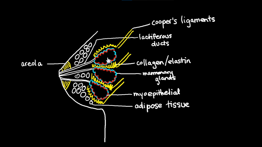
Breast fat is an important part of women’s breast Anatomy. This fat tissue is located in and around the mammary glands and detects the shape, contour, and size of a woman’s breasts. The adipose tissue of the mammary gland not only affects the formation of female breasts, but also affects many other biological functions, such as breastfeeding and hormonal regulation.
The fatty tissues of women’s breasts are made up of lobular cells, which are the same as the mammary glands, and stromal cells that make up the main breast structure.
This type of fat is structurally designed for our body shape and helps perform functions such as providing shape and definition to our body lines. Plus it helps us to do our daily activities easily. In addition, it regulates the temperature inside and helps to maintain heat and insulate the environment during the colder months.
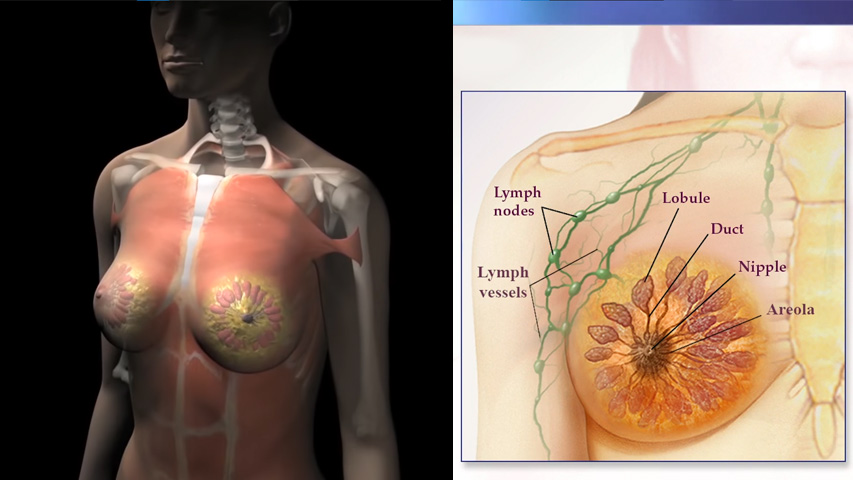
Lymph nodes and vessels
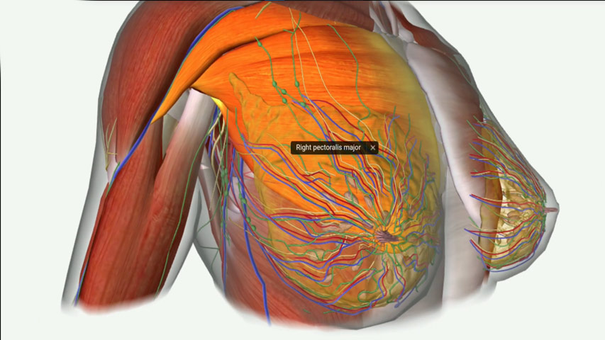
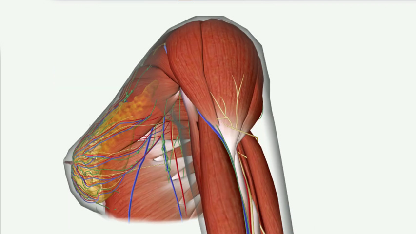
The anatomy of Women’s Pectoral Muscles includes various organs, tissues and nodules that play an important role. These nodes include the lymph nodes and thoracic veins, which play an important role in the circulation of fluids in the body and regulating blood circulation.
Lobe and Lobules
As part of the recognition of breast Anatomy, important structures such as lobes and lobules in the breast are responsible for the production of milk. Lobes are small, rounded specialized glands that contain milk-producing cells called alveoli, while lobules are ways of communicating between them.
Lobules are masses or small groups of these Alveoli that can produce small amounts of milk at any time. The mammary glands in this area provide nutrients for infants.
It is good to know that postpartum breastfeeding has become an important issue in most countries of the world !
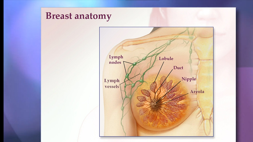
Diagram of Female Breast Muscles
The anatomy of the female pectoral muscles is such that each breast has 15 to 20 lobes and is arranged like daisies. Each lobe consists of smaller structures called lobules. They end up with dozens of tiny lamps that can produce milk. The lobes and bulbs are connected by thin tubes called ducts. These tubes lead to the nipple in the center of the dark area of the skin called areol.
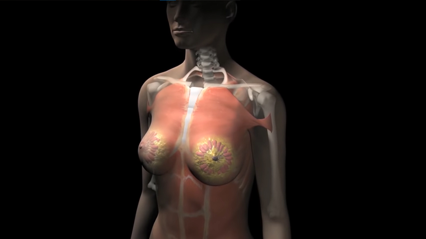
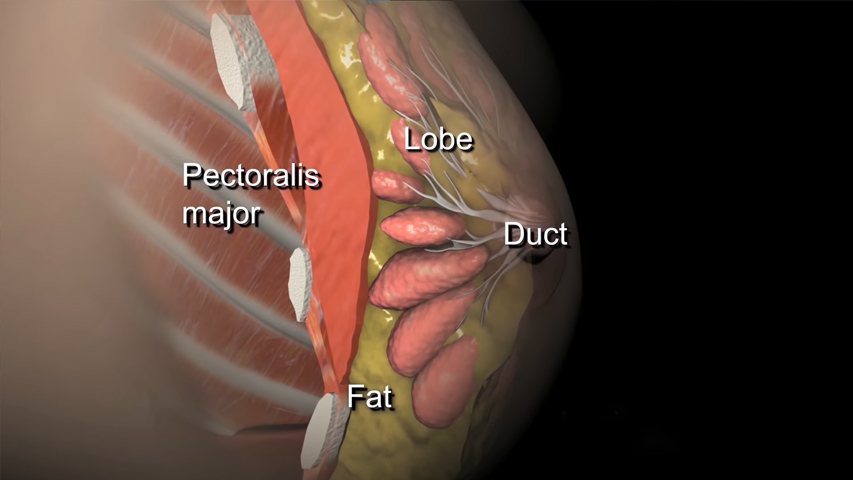
There are no muscles in the chest, but there are muscles under each breast that cover the ribs. Each breast also contains blood vessels and vessels that carry the lymph nodes. You can better understand the anatomy of the pectoral muscles according to the chart of women’s pectoral muscles. Here is a diagram of the anatomy of the pectoral muscles.
Women Chest Muscles Anatomy
Breasts are double structures located in the superficial fascia of the posterior wall of the chest. Real breast development in women begins at puberty and about 10 years of age under the influence of estrogen and progesterone. Before puberty, both in women and men, the mammary glands consist of only a few ducts in the stroma of connective tissue. Women also have mammary glands in their breasts.
Breast Glands are one of the main concerns of breastfeeding. The mammary gland is also an important part of the female reproductive system. The anatomy of the pectoral muscles of women is present in various types such as superficial Anatomy, menstrual period Anatomy and menopausal anatomy of women, etc. We’re going to look at each of them here.
1) Superficial Anatomy of Women’s Breasts
The chest is in the form of a Hemisphere with an extension towards the armpits, and with age, it becomes more sagging. The chest continues from the surface of the second rib in the middle of the clavicle line to the seventh rib and on muscles that extend from the outer edge of the sternum to the middle of the underarm line. It is located on the pectoral muscles of the major, anterior and right abdominal ceratus. The breast has 12 to 15 main milk ducts that drain into the nipple.
2) Anatomy of Breast tissue during Menstruation
Menstruation is an important part of a woman’s life. The hormones involved in this process can cause many changes, including changes in the anatomy and breast tissue, in women’s bodies. During menstruation, an increase in Hormones such as estrogen and progesterone can cause swelling, tenderness or lumping of the breasts. This increase in hormones can lead to severe discomfort or pain.
3) Breast Anatomy during menopause
During menopause, many changes occur in the female body, such as hormonal imbalances, changes in the anatomy of the breast, and loss of fat cells. The severity of these changes can be more or less, depending on the person. Menopause is a natural process in every woman’s life, and the changes that occur in her body are very noticeable. One of the organs that undergoes a lot of changes after menopause is the anatomy of women’s chest muscles.
These changes include thinning of the skin around the nipples, loss of breast tissue, and accumulation of fat near the armpits. Also during menopause, women experience tenderness and chest pain due to hormonal changes, as well as physical symptoms of smaller breasts and loss of stiffer breasts. Aware of these changes, women can take better care of themselves, recognize early warning signs in a timely manner and refer them to a specialist for treatment.
Female breast Muscles
Pectoral muscles are muscles that connect the front of the breast to the shoulder and arm bones. The muscles of the breasts of women include:
- Deep pectoral muscles that include pectoralis minor, anterior ceratus, and subclavianis.
- The major pectoral muscle is a thick sail-shaped muscle that forms the majority of the pectoral muscle and is located under the pectoral. The long bone carries the arm in a state of stretching, bending and rotation.
- The minor pectoral muscle is a thin triangular-shaped muscle located deep in the thoracic muscle. It adheres to the ribs and stabilizes the large shoulder bone, the shoulder blade.
- The pectoral Niamh muscle is a thin layer of tissue on the large pectoral muscle that extends to the large pectoral muscle at the back.
- The subclavian muscle together with the large pectoral and pectoral muscles forms the minor underarm. The muscle under the clavicle moves the shoulder forward and down.
- The Anterior tooth Muscle is another muscle in front of the chest that moves the shoulder forward around the trunk.
- The intercostal muscles that are present between the ribs and help with breathing.
last Word about Women Chest Muscles Anatomy
One way to succeed in bodybuilding and become an expert in this field is to fully understand muscle anatomy. Because after you’re fully familiar with The Shape of your muscles, you can do your workout routine in a principled and professional way and improve as quickly as possible. Breast is the most common cancer case in women, knowing the anatomy of women’s breast muscles can help us understand the different types of breast cancer and their origin, diagnosis and treatment. Studying breast anatomy can be very helpful in early detection of any abnormalities that may develop in the future or cause health problems.
healthline